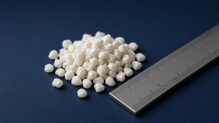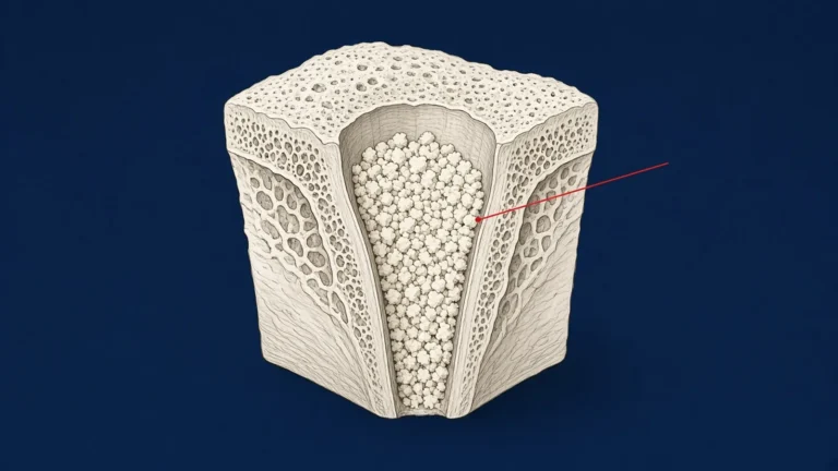
Bone graft materials
Bone graft materials in dentistry: autograft, allograft, xenograft, alloplast and tooth-derived
How the literature defines the five material categories, the three biological properties used to compare them, and what systematic reviews of ridge preservation found when they compared categories.
Vol. 1No. 013 min read5 references
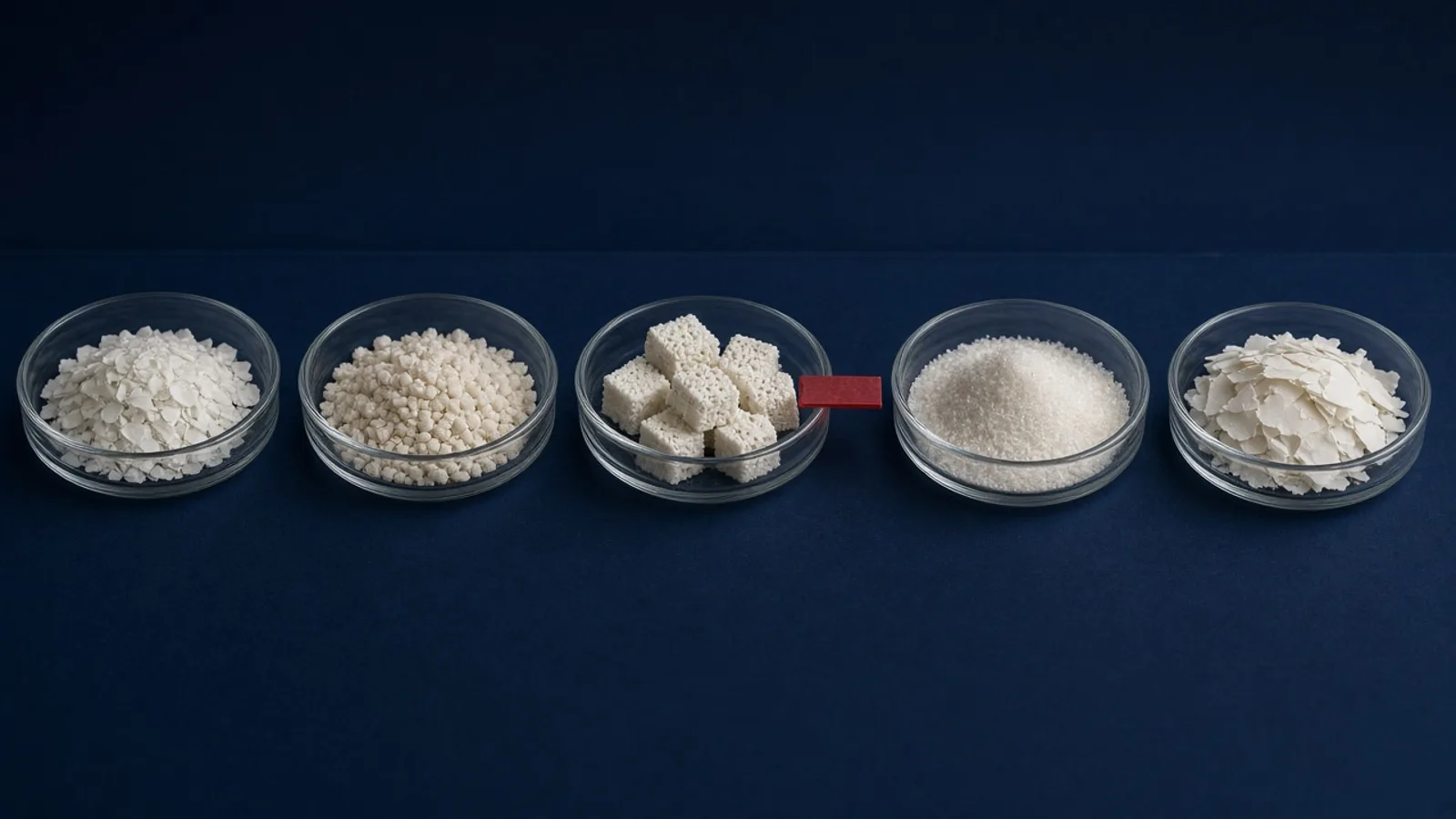
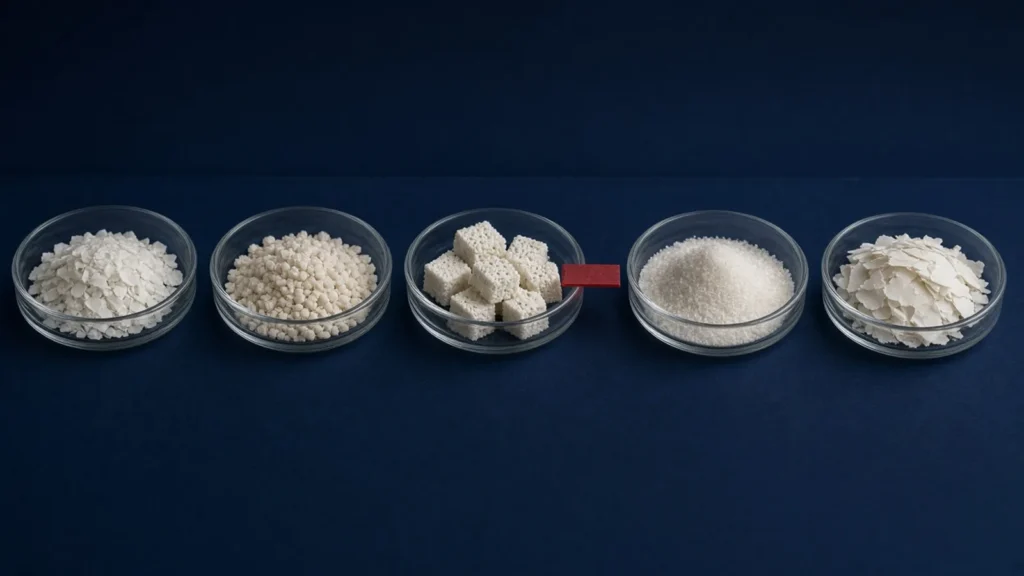
Bone graft materials are usually grouped by where they come from: the patient, another person, an animal, a laboratory, or a tooth. This article sets out how the published literature defines each group, and what systematic reviews found when they compared groups in ridge preservation. It is a reference for clinicians. It does not recommend a product.
Three properties used to compare grafts
Most reviews compare graft materials on three biological properties. In the definitions used by Ferraz (2023):1
Source 1
- 1 Ferraz MP, 2023. Bone Grafts in Dental Medicine: An Overview of Autografts, Allografts and Synthetic Materials
- Osteogenic materials contain living cells that can differentiate into bone.
- Osteoinduction is the process by which new bone formation is induced from osteocompetent cells.
- Osteoconduction is bony ingrowth from local osseous tissue onto the graft surface.
A material can have one, two or all three. The categories below differ mainly in which of these they are described as having.
Autografts: the patient’s own bone
Autologous bone is taken from the patient, for example from the iliac crest or the calvaria for large defects. The review describes it as the reference material for bone grafting because it is biocompatible, osteogenic, osteoinductive and osteoconductive.1 Its drawbacks are the need for surgery at a donor site, limited availability and an uncertain prognosis.1
Allografts: human bone from another person
Allografts are human bone from a donor other than the patient, selected and processed before use. Ferraz describes them as osteoinductive and osteoconductive but not osteogenic. The same review notes continuing debate about immune response and disease transmission, and that processing reduces some biological and mechanical characteristics.1
Xenografts: bone from another species
Xenografts are bone from animals. According to the review, their osteoconductivity comes from the inorganic structure, mainly hydroxyapatite, left after organic components are removed. Their osteoinductive capacity is described as questionable when no organic component remains.1
Alloplasts: synthetic materials
Synthetic materials include ceramics, bioactive glasses, polymers and synthetic hydroxyapatite. The review lists biocompatibility, osteoconduction and easy handling among their advantages. It also notes that calcium phosphates may lack osteoinductivity, which restricts their use in larger defects.1
Tooth-derived grafts
Grafts can also be made from dentin. Solyom and colleagues (2023) describe three types by degree of demineralization: undemineralized dentin matrix (UDDM), partially demineralized dentin matrix (PDDM) and demineralized dentin matrix (DDM).4 Most published clinical studies in this group used the patient’s own extracted teeth (autogenous tooth grafts). Our article on tooth-derived bone grafts covers this group in more detail.
Source 4
- 4 Solyom E et al, 2023. The use of autogenous tooth bone graft is an efficient method of alveolar ridge preservation – meta-analysis and systematic review
AmGraft® belongs to this broad family. Its Instructions for Use describe it as “a dentin matrix, which is made from human teeth”, supplied as chips of 500–1000 microns, freeze-dried and sterile.5 It is a packaged product, not a chairside preparation of the patient’s own tooth. The autogenous studies cited in this article are therefore not studies of AmGraft.
Source 5
- 5 Ventas Bio Pte Ltd, 2022. AmGraft® Instructions for Use (date of information 2022-06)
Does the category change ridge preservation outcomes?
Two systematic reviews of alveolar ridge preservation (ARP) compared material categories.
- Majzoub et al. (2019) pooled 40 randomised controlled trials. Average horizontal ridge resorption was 1.52 mm with allograft, 1.47 mm with xenograft, 2.31 mm with alloplast and 3.1 mm with unassisted healing. The authors concluded that grafting reduced resorption after extraction, and that differences between allogeneic, xenogeneic and alloplastic materials were minimal.2
- The 2021 Cochrane review (Atieh et al.) found that ARP techniques may reduce changes in ridge height and width six months after extraction, but rated the evidence as very uncertain. It found no evidence of a clinically significant difference between grafting materials or barriers used for ARP.3
Sources 2, 3
- 2 Majzoub J et al, 2019. The Influence of Different Grafting Materials on Alveolar Ridge Preservation: a Systematic Review
- 3 Atieh MA et al, 2021. Interventions for replacing missing teeth: alveolar ridge preservation techniques for dental implant site development
Read together, these reviews suggest that grafting itself matters more for ridge dimensions than the choice of material category. Long-term outcome data, such as implant failure and aesthetics, remain limited.3
Choosing a material
Ferraz lists the factors that drive selection: material availability, defect size, the size, shape and volume of graft needed, biomechanics, handling, cost, ethical issues, biological characteristics and associated complications.1 Two practical points follow:
- Check the indication list. Every graft is cleared or notified for specific indications. Use it only within the indications in its Instructions for Use.
- Read the precautions and the healing time. These vary between products. For example, the AmGraft IFU states that the product is not intended to bear mechanical loads and recommends a minimum of 6 months’ healing at the augmented site.5
Clinicians who want AmGraft’s full IFU-based reference (indications, contraindications, precautions and handling) can find it in the professional area.
References
- 1Ferraz MP. Bone Grafts in Dental Medicine: An Overview of Autografts, Allografts and Synthetic Materials. Materials (Basel). 2023;16(11):4117. doi:10.3390/ma16114117
- 2Majzoub J, et al. The Influence of Different Grafting Materials on Alveolar Ridge Preservation: a Systematic Review. J Oral Maxillofac Res. 2019;10(3):e6. doi:10.5037/jomr.2019.10306
- 3Atieh MA, et al. Interventions for replacing missing teeth: alveolar ridge preservation techniques for dental implant site development. Cochrane Database Syst Rev. 2021;(4):CD010176. doi:10.1002/14651858.CD010176.pub3
- 4Solyom E, et al. The use of autogenous tooth bone graft is an efficient method of alveolar ridge preservation – meta-analysis and systematic review. BMC Oral Health. 2023;23:226. doi:10.1186/s12903-023-02930-2
- 5Ventas Bio Pte Ltd. AmGraft® Instructions for Use (date of information 2022-06).
- allograft
- alloplast
- autograft
- ridge preservation
- xenograft
ShareCopy linkEmail a colleagueLinkedIn
General education for dental professionals. Not advice on the care of any individual patient. Statements about AmGraft® follow its Instructions for Use (2022-06). AmGraft® is manufactured by Ventas Bio Pte Ltd and distributed by Santé Maison.