Clinical practice
Socket preservation after extraction: what the evidence supports
Ridge preservation reduces post-extraction bone loss in most trials, but the certainty of evidence and the long-term benefit are debated. A summary of the reviews and the consensus timing guidance.
Vol. 1No. 023 min read5 references
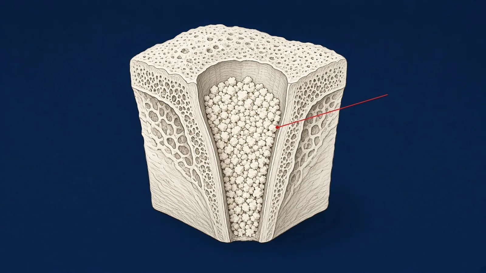
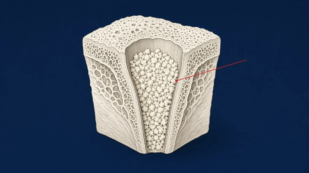
After a tooth is extracted, the ridge changes shape as the socket heals. Alveolar ridge preservation (ARP) is an intervention at the time of extraction intended to limit those changes. This article summarises what systematic reviews and the 2019 European Workshop in Periodontology consensus say about it.
What counts as ridge preservation
The European Workshop’s review defined ARP interventions as filling the socket with a biomaterial (socket grafting), sealing the socket with a barrier material alone, or a combination of both, with either primary or secondary intention healing.2 This article focuses on socket grafting.
Source 2
- 2 Tonetti MS et al, 2019. Management of the extraction socket and timing of implant placement: Consensus report and clinical recommendations of group 3 of the XV European Workshop in Periodontology
How much bone does it preserve?
The consensus report states, at its highest strength of statement, that socket grafting attenuates the dimensional changes that typically follow extraction. Compared with extraction alone, it may prevent 1.5–2.4 mm of horizontal resorption, 1–2.5 mm of mid-buccal vertical resorption and 0.8–1.5 mm of mid-lingual vertical resorption.2
A separate review of 40 randomised trials reported average horizontal resorption of 3.1 mm with unassisted healing, against 1.47–2.31 mm in grafted groups depending on material category.3
Source 3
- 3 Majzoub J et al, 2019. The Influence of Different Grafting Materials on Alveolar Ridge Preservation: a Systematic Review
How certain is that evidence?
The reviews differ in how confident they are.
- The Cochrane review (2021) concluded that ARP techniques may minimise changes in ridge height and width six months after extraction, but that the evidence is very uncertain. It found a lack of evidence for differences in the need for additional augmentation, implant failure or aesthetic outcomes, and no evidence of a clinically significant difference between grafting materials or barriers.1
- Fischer et al. (2022) looked at whether ARP changes the bone available for implant placement. Using deproteinised bovine bone mineral, ARP reduced dimensional change by 1.13 mm on average. On the authors’ calculation, roughly one patient in five would benefit, so the clinical benefit “remains unclear”.4
- The consensus report found that implant placement without simultaneous grafting is more feasible after socket grafting, but that additional augmentation may still be needed after either approach.2
Sources 1, 4
- 1 Atieh MA et al, 2021. Interventions for replacing missing teeth: alveolar ridge preservation techniques for dental implant site development
- 4 Fischer KR et al, 2022. Bone envelope for implant placement after alveolar ridge preservation: a systematic review and meta-analysis
Healing time before implant placement
The consensus report recommends a minimum healing time that allows sufficient bone formation, typically 3–4 months. It adds that a longer healing time may be needed depending on the phenotype of the extraction site, the properties of the biomaterial used, and systemic factors specific to the patient.2
The biomaterial’s own Instructions for Use therefore matter. For example, the AmGraft® IFU recommends a minimum of 6 months’ healing at the augmented site and leaves the final decision to the practitioner.5 Our article on healing time after grafting looks at this in more detail.
Source 5
- 5 Ventas Bio Pte Ltd, 2022. AmGraft® Instructions for Use (date of information 2022-06)
Where AmGraft’s IFU applies
“Filling of alveolar defects following dental extraction” is one of the five clinical uses listed in the AmGraft IFU.5 The IFU’s handling instructions for any site include:
- Remove bone fragments, granulation tissue and necrotic tissue carefully before applying the graft.
- Place the chips directly, or hydrate or mix them with the patient’s blood from the surgical site or with saline.
- The mucoperiosteal flaps can be sutured to achieve primary closure and minimise particle loss. In some cases the surgeon may want to place a surgical dressing or membrane over the wound.
The IFU contraindicates use where general oral surgery is contraindicated, for example infection at the grafting site. It also lists conditions and therapies that call for special caution, including poorly controlled diabetes, radiotherapy and heavy smoking.5
The full IFU text for this indication is in the professional area.
References
- 1Atieh MA, et al. Interventions for replacing missing teeth: alveolar ridge preservation techniques for dental implant site development. Cochrane Database Syst Rev. 2021;(4):CD010176. doi:10.1002/14651858.CD010176.pub3
- 2Tonetti MS, et al. Management of the extraction socket and timing of implant placement: Consensus report and clinical recommendations of group 3 of the XV European Workshop in Periodontology. J Clin Periodontol. 2019. doi:10.1111/jcpe.13131
- 3Majzoub J, et al. The Influence of Different Grafting Materials on Alveolar Ridge Preservation: a Systematic Review. J Oral Maxillofac Res. 2019;10(3):e6. doi:10.5037/jomr.2019.10306
- 4Fischer KR, et al. Bone envelope for implant placement after alveolar ridge preservation: a systematic review and meta-analysis. Int J Implant Dent. 2022;8:56. doi:10.1186/s40729-022-00453-z
- 5Ventas Bio Pte Ltd. AmGraft® Instructions for Use (date of information 2022-06).
- ridge preservation
- socket preservation
- tooth extraction
ShareCopy linkEmail a colleagueLinkedIn
General education for dental professionals. Not advice on the care of any individual patient. Statements about AmGraft® follow its Instructions for Use (2022-06). AmGraft® is manufactured by Ventas Bio Pte Ltd and distributed by Santé Maison.