Tooth-derived grafts
Tooth-derived bone grafts explained: what the autogenous dentin studies show
Two meta-analyses of autogenous dentin grafts in ridge preservation, how dentin grafts are classified, and why results from a patient’s own tooth cannot be read across to a manufactured product.
Vol. 1No. 033 min read3 references
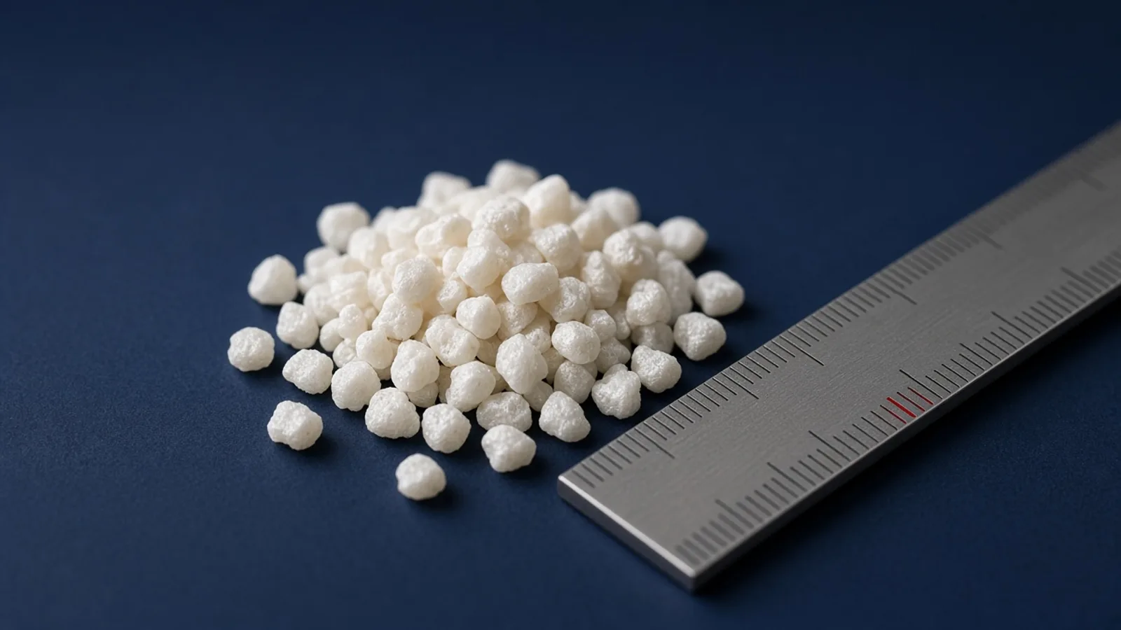
Dentin and bone are both mineralised tissues, so extracted teeth have been studied as a source of graft material. This article explains how dentin grafts are classified and what two recent meta-analyses found. It also makes one distinction clear: the published clinical studies used autogenous tooth grafts, made from the patient’s own extracted tooth. They are not studies of AmGraft®, and their results should not be read as AmGraft results.
How autogenous tooth grafts are made
Several protocols have been proposed for making a graft from a patient’s extracted tooth. They commonly involve removing soft tissue, carious lesions and fillings after extraction.1 The tooth is then processed into particles.
Source 1
- 1 Solyom E et al, 2023. The use of autogenous tooth bone graft is an efficient method of alveolar ridge preservation – meta-analysis and systematic review
Three types, by degree of demineralization
Solyom and colleagues describe three main types:1
- Undemineralized dentin matrix (UDDM)
- Partially demineralized dentin matrix (PDDM)
- Demineralized dentin matrix (DDM)
The degree of demineralization appears to matter. In their pooled data, the proportion of newly formed bone was highest in the PDDM group (51%) and lowest in the DDM group (31%). The authors concluded that complete demineralization tends to decrease the proportion of new bone.1
What the meta-analyses found
Solyom et al., 2023
This systematic review pooled eight studies (96 patients) for ridge width and six studies (103 patients) for histology, measured 3–6 months after ridge preservation.1
- Pooled ridge width change: −0.72 mm.
- Residual graft: 11.61% on average.
- Newly formed bone: 40.23% on average.
The authors called autogenous tooth bone graft an effective particulate material for ridge preservation. They also noted that earlier clinical studies had small samples, so conclusions “rely on weak evidence”, and that measurement methods varied between the studies they pooled.1
Zhang et al., 2025
This network meta-analysis included 25 randomised controlled trials with 710 patients and 830 extraction sites. It compared autologous particulate dentin with xenogeneic, allogeneic and alloplastic grafts and with blood-clot healing.2
Source 2
- 2 Zhang Y et al, 2025. Comparative effectiveness of autologous particulate dentin graft for alveolar ridge preservation: a network meta-analysis of randomized controlled trials
- Compared with blood-clot healing, autologous dentin preserved 1.67 mm more coronal ridge width.
- Compared with xenograft, the difference in coronal width was 0.75 mm in favour of dentin.
- Both dentin and xenograft maintained buccal and lingual ridge height better than blood-clot healing.
The authors rated confidence in these comparisons as moderate to low. They stressed that several comparisons relied on indirect evidence and that network rankings “are best viewed as comparative summaries rather than definitive statements of superiority”. They recommended that material choice be individualised by handling, availability, preference, cost, timing and experience.2
Why these results do not transfer to AmGraft
Autogenous tooth grafts are prepared from the patient’s own tooth, usually at the time of extraction. AmGraft is a different kind of product. Its Instructions for Use describe it as “a dentin matrix, which is made from human teeth”, produced as chips of 500–1000 microns and supplied freeze-dried and sterile in sealed packaging.3
Source 3
- 3 Ventas Bio Pte Ltd, 2022. AmGraft® Instructions for Use (date of information 2022-06)
Source tissue, processing and form all differ from the autogenous protocols studied above. So the figures in this article describe autogenous dentin only. They are not evidence about AmGraft’s performance, and they should not be quoted as such.
For AmGraft, the IFU is the reference for intended use, indications, contraindications, precautions and handling.3 The manufacturer’s own laboratory and animal data are summarised in the professional area, with each item labelled by evidence type (in vitro or preclinical animal study, data on file).
Practical points
- When you read about dentin grafts, check whether the study used autogenous tooth material, the degree of demineralization, and the healing interval measured.
- Read each product’s IFU for its indications and healing time. The AmGraft IFU, for example, recommends at least 6 months’ healing at the augmented site and states that the product is not intended to bear mechanical loads.3
References
- 1Solyom E, et al. The use of autogenous tooth bone graft is an efficient method of alveolar ridge preservation – meta-analysis and systematic review. BMC Oral Health. 2023;23:226. doi:10.1186/s12903-023-02930-2
- 2Zhang Y, et al. Comparative effectiveness of autologous particulate dentin graft for alveolar ridge preservation: a network meta-analysis of randomized controlled trials. BMC Oral Health. 2025;25:1914. doi:10.1186/s12903-025-07371-7
- 3Ventas Bio Pte Ltd. AmGraft® Instructions for Use (date of information 2022-06).
- autogenous dentin
- ridge preservation
- tooth-derived graft
ShareCopy linkEmail a colleagueLinkedIn
General education for dental professionals. Not advice on the care of any individual patient. Statements about AmGraft® follow its Instructions for Use (2022-06). AmGraft® is manufactured by Ventas Bio Pte Ltd and distributed by Santé Maison.